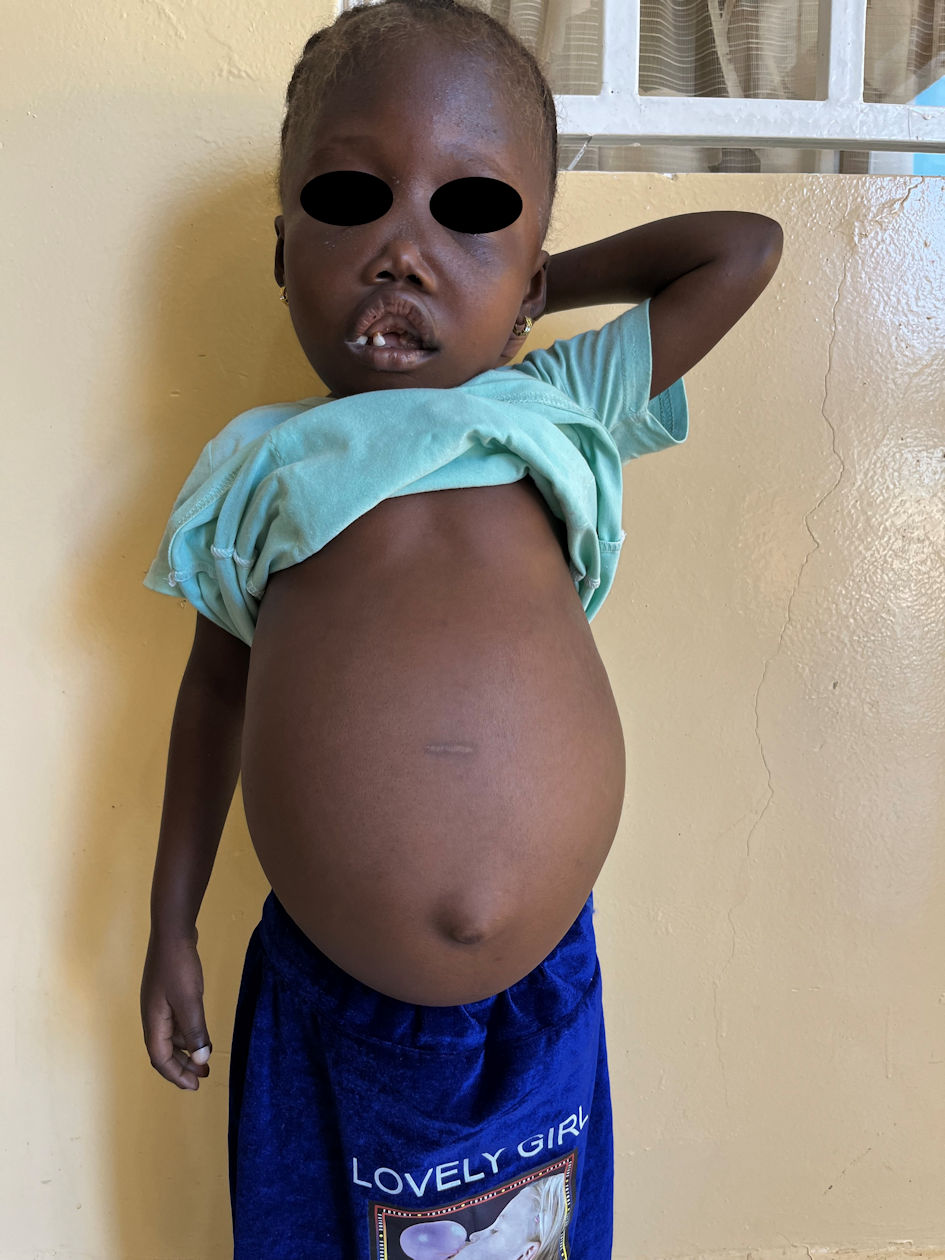
↓ Figure 1. Clinical image. Note the marked
pallor, prominent forehead, flat nasal bridge, marked prominence of the maxilla, gingival hypertrophy
with disordered dentition and grossly distended abdomen.
| International Journal of Clinical Pediatrics, ISSN 1927-1255 print, 1927-1263 online, Open Access |
| Article copyright, the authors; Journal compilation copyright, Int J Clin Pediatr and Elmer Press Inc |
| Journal website https://ijcp.elmerpub.com |
Case Report
Volume 14, Number 2, October 2025, pages 51-54
A Case of Homozygous Beta+-Thalassemia
Figures
Table
| Patient | Father | |
|---|---|---|
| Normal reference ranges are shown in parentheses. Hb: hemoglobin; HbF: fetal hemoglobin; HbA: adult hemoglobin. | ||
| Hb concentration (g/dL) | 6.1 (11.5 - 14.0) | 13.8 (14.0 - 18.0) |
| Mean cell volume (fL) | 61 (80 - 100) | 64.1 (80 - 100) |
| Mean cell Hb (pg) | 16.1 (27.0 - 31.0) | 22.2 (27.0 - 31.0) |
| Red blood cell count (1012/L) | 2.3 (4.0 - 5.5) | 6.2 (4.0 - 5.5) |
| Reticulocytes (%) | 10.7 (0.4 - 3.0) | - |
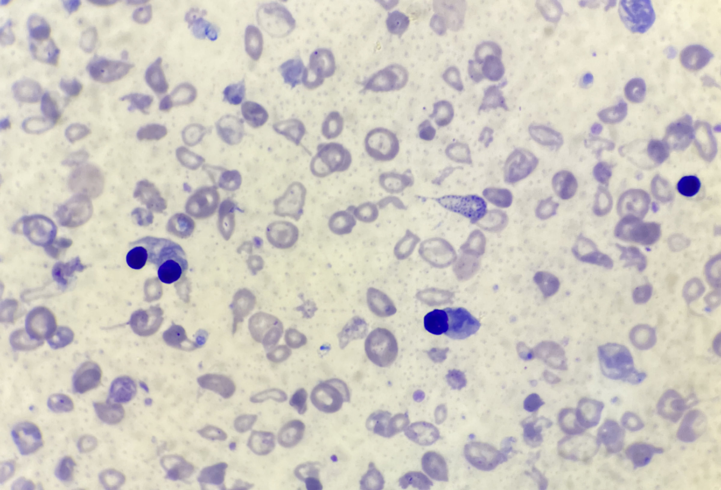
| Hb electrophoresis | Predominantly HbF, with some HbA and a raised HbA2 | Predominantly HbA with a raised HbA2 |
| HbA2 (%) | 4.5 (2.5 - 3.1) | 4.1 (2.5 - 3.1) |
| White blood cell count (109/L) | 75.8 (4.5 - 11.0) | - |
| White blood cell differential (%) | Lymphocytes 68.3% (20 - 40); granulocytes 25.5 (41.5 - 68.0) | - |